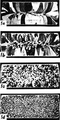
Photograph 1 shows the crystals in aluminum ingots with different additions of Ti. In picture 1a there is no Ti and no nuclei have formed inside the ingot. All crystals have nucleated at the surface and grown inwards in a columnar fashion. The Ti content has then been increased to 0.03, 0.06 and 0.10 % in 1b, 1c and 1d. The number of crystals nucleated at the surface has thus increased and new nuclei appear in a central region of the ingots. They appear earlier the higher the Ti content is. Within the central region the new nuclei seem to be randomly distributed and give rise to equiaxed crystals (crystals with approximately the same width in different directions). One often talks about the columnar zone and the equiaxed zone.
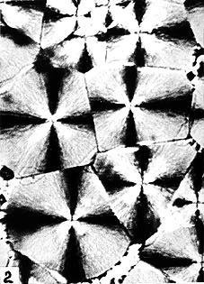
Micrograph 2 shows the structure of a crystallized polymer. The crystallization starts from nuclei and proceeds with the same rate in all directions. Each unit is thus spherical during growth until they impinge on each other (so-called hard impingement) and get a polyhedral shape. The spherical shape can still be seen at the lower left part of the picture where the crystallization was not completed when interrupted by cooling. The picture was taken with polarized light and the crosses reveal the positions of the polarizer and analyzer.
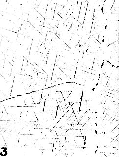
Micrograph 3 shows a case where nuclei have formed at random inside the grains of a solid Al alloy and each nucleus has grown into a plate-like crystal (so-called WidmanstŠtten plates). The growth of each crystal has stopped when it was approaching other crystals, actually when it was entering the depleted zone around other crystals (so-called soft impingement). Their actual shape during growth is preserved rather well.
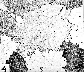
Micrograph 4 shows a solidified Al alloy where the nuclei were distributed at random. They have grown with almost the same rate in all directions until impinging on each other. In contrast to picture 2, the final crystal boundaries are here very jagged and reveal that the crystals were actually dendritic during growth although they became compact when the solidification was completed.
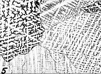
Micrograph 5 shows a similar case in a solidified Cu alloy. Here the etching method reveals a dendritic pattern reflecting the shape at an early stage of solidification.